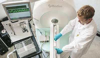
Positron emission tomography (PET)
Positron emission tomography
Positron emission tomography (PET) is a functional imaging technique that uses radioactive substances known as radiotracers to visualize and measure changes in metabolic processes, and in other physiological activities including blood flow, regional chemical composition, and absorption. In clinical practice it is used to diagnose and manage cancer treatment, in cardiology and cardiac surgery, and in neurology and psychiatry. PET is a common imaging technique, a medical scintillography technique used in nuclear medicine. A radiopharmaceutical—a radioisotope attached to a drug—is injected into the body as a tracer. When the radiopharmaceutical undergoes beta plus decay, a positron is emitted, and when the positron interacts with an ordinary electron, the two particles annihilate and two gamma rays are emitted in opposite directions. These gamma rays are detected by two gamma cameras to form a three-dimensional image. PET scanners can incorporate a computed tomography scanner (CT) and are known as PET–CT scanners. PET scan images can be reconstructed using a CT scan performed using one scanner during the same session. One of the disadvantages of a PET scanner is its high initial cost and ongoing operating costs.
Read more about 'Positron emission tomography' at: WikipediaWikipedia contributors. "Positron emission tomography." Wikipedia, The Free Encyclopedia. Wikipedia, The Free Encyclopedia, Feb. 13, 2026.
Positron emission tomography (PET) is a unique modality for imaging normal and diseased metabolical functions in vivo, in a quantitative manner and is a type of nuclear medicine imaging. It is a functional imaging technique that uses radioactive substances known as radiotracers to visualize and measure changes in metabolic processes, and in other physiological activities including blood flow, regional chemical composition, and absorption. Different tracers are used for various imaging purposes, depending on the target process within the body. Radiotracers are molecules linked to, or "labeled" with, a small amount of radioactive material. They accumulate in tumors or regions of inflammation. They can also bind to specific proteins in the body.
At Helmholtz-Zentrum Dresden-Rossendorf the in-beam Positron-Emission-Tomography (PET) has been developed as a technology for the visualizatio of a radiation field in ion beam therapy. The objective of this method is the verification of the correct dose application to the target volume and the exclusion of an unwanted irradiation of healthy tissue.
** Resources used **
"Positron emission tomography." Wikipedia, The Free Encyclopedia. Wikipedia, The Free Encyclopedia, 5 Feb. 2022. Web. 8 Feb. 2022. RadiologyInfo - Positron Emission Tomography - Computed Tomography (PET/CT)Positron-Emission-Tomography for ion beam therapy (PT-PET)
See also:
Experimental and Computational Methods at Institute of Resource Ecology
Department of Positron Emission Tomography at HZDR
Department of Neuroradiopharmaceuticals: project Flubatine, project VAChT, CB2 project, PDE10, project Sigma 1
