Joint Inversion for Accurate Brain Myelin Mapping (JIMM)
Keywords: myelin, myelin water imaging, qMRI
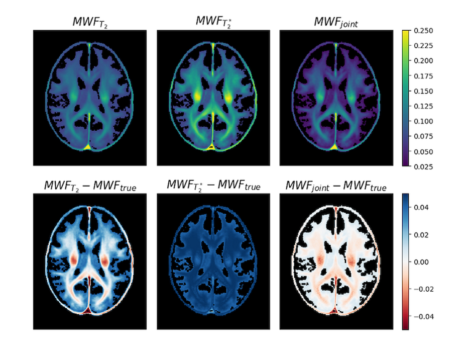
Magnetic Resonance Imaging (MRI) is a routine tool in clinical diagnostics. In the past MRI was mainly used for subjective image reading by experienced radiologists, but the technology can be extended to quantitative biophysical tissue parameter mapping. Sensitizing the acquisition to the process of interest enables parameter estimation by subsequent model fitting of the MRI data. An important example is the estimation of myelin content. Myelin is a major factor for brain structure and function and brain demyelination is a consequence of many neurological diseases. Since myelin affects many physical processes, there exist different MRI-based approaches for myelin quantification. These approaches are based on model simplifications and assumptions, which result in biased estimates of myelin content. The idea of JIMM is to simultaneously fit two or more of these models by using joint inversion approaches well-known from geophysical imaging. Due to the inherently ill-posed nature of geophysical inverse problems, there exist long experience and a variety of mathematical tools to combine data of different physical nature to fit a single or even different material parameters. Such methodology was not yet applied in medical imaging applications. Joint inversion improves accuracy and reproducibility of myelin mapping as compared to conventional approaches.
Publications
Myelin water fraction mapping with joint inversion of gradient-echo and spin-echo data
Dega S, Ferreira M, Veldmann M, Stirnberg R, Paasche H, Stöcker T - Magnetic Resonance Materials in Physics, Biology and Medicine - 2025
Helmholtz Imaging Projects
2020: JIMM - Geophysical Joint Inversion for Accurate Brain Myelin Mapping