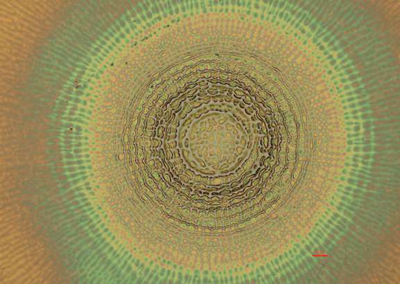
Bright field imaging
Bright-field microscopy
Bright-field microscopy (BF) is the simplest of all the optical microscopy illumination techniques. Sample illumination is transmitted (i.e., illuminated from below and observed from above) white light, and contrast in the image is caused by attenuation of the transmitted light in dense areas of the sample. Bright-field microscopy is the simplest of a range of techniques used for illumination of samples in light microscopes, and its simplicity makes it a popular technique. The typical appearance of a bright-field microscopy image is a dark sample on a bright background, hence the name.
Read more about 'Bright-field microscopy' at: WikipediaWikipedia contributors. "Bright-field microscopy." Wikipedia, The Free Encyclopedia. Wikipedia, The Free Encyclopedia, Dec. 8, 2025.
Bright-field microscopy, a method of microscopy in which the object to be observed is located in front of a bright background, called the bright field, which illuminates the microscopic image. In this process, the light passing through the object (transmitted light microscopy) or the light reflected back from it as a whole (reflected light microscopy) enters the objective of the microscope. Bright-field microscopy requires high-contrast objects, so many transmitted-light objects must be stained. In brightfield microscopy, objects appear dark or colored on a light background.
Resources used
