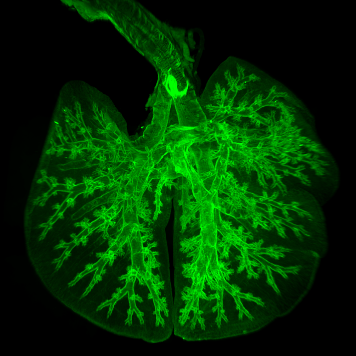
Light sheet fluorescence microscopy (LSFM)
Light sheet fluorescence microscopy
Light-sheet fluorescence microscopy (LSFM) is a fluorescence microscopy technique with an intermediate-to-high optical resolution, but good optical sectioning capabilities and high speed. In contrast to epifluorescence microscopy only a thin slice (usually a few hundred nanometers to a few micrometers) of the sample is illuminated perpendicularly to the direction of observation. For illumination, a laser light-sheet is used, i.e. a laser beam which is focused only in one direction (e.g. using a cylindrical lens). A second method uses a circular beam scanned in one direction to create the light-sheet. As only the actually observed section is illuminated, this method reduces the photodamage and stress induced on a living sample. Also the good optical sectioning capability reduces the background signal and thus creates images with higher contrast, comparable to confocal microscopy. Because light-sheet fluorescence microscopy scans samples by using a plane of light instead of a point (as in confocal microscopy), it can acquire images at speeds 100 to 1,000 times faster than those offered by point-scanning methods. This method is used in cell biology and for microscopy of intact, often chemically cleared, organs, embryos, and organisms. Starting in 1994, light-sheet fluorescence microscopy was developed as orthogonal plane fluorescence optical sectioning microscopy or tomography (OPFOS) mainly for large samples and later as the selective/single plane illumination microscopy (SPIM) also with sub-cellular resolution. This introduced an illumination scheme into fluorescence microscopy, which has already been used successfully for dark-field microscopy under the name ultramicroscopy.
Read more about 'Light sheet fluorescence microscopy' at: WikipediaWikipedia contributors. "Light sheet fluorescence microscopy." Wikipedia, The Free Encyclopedia. Wikipedia, The Free Encyclopedia, Sept. 25, 2026.
Light Sheet Microscopy is a special fluorescence microscopic technique in which only a thin layer in the sample is illuminated, typically a few micrometers. Compared to conventional fluorescence microscopy, this results in better resolution and significantly reduced background signal. In addition, negative effects due to bleaching or light-induced stress on a living sample are reduced.