Photoacoustic Imaging
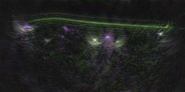
When a laser beam is shined into tissue, the absorption of light causes a thermal expansion of the surrounding tissue, which in turn generates a shock wave. This phenomenon is known as the photoacoustic effect. The resulting shock waves can be measured with an ultrasound probe, allowing insights into the tissue composition. Due to their high absorption coefficient, hemoglobin and melanin distributions are popular targets for this imaging modality. Facilitated by differences in absorption spectra between the oxygenated and deoxygenated states of hemoglobin, blood oxygenation detection is one of the most popular applications of photoacoustic imaging.
There are two major imaging methods making use of the photoacoustic effect. In photoacoustic computed tomography, an unfocused detector is used and the image must be reconstructed by inversely solving photoacoustic equations. In photoacoustic tomography, a focused ultrasound detector is used which uses point-by-point rasterization.
Resources used
Publications
A review of clinical photoacoustic imaging: Current and future trends
Attia A, Balasundaram G, Moothanchery M, Dinish U, Bi R, Ntziachristos V, Olivo M - Photoacoustics - 2019
Photoacoustic Imaging
Zhang Y, Hong H, Cai W - Cold Spring Harbor Protocols - 2011
Photoacoustic imaging and characterization of the microvasculature
Hu S, Wang L - Journal of Biomedical Optics - 2010
